University of Tokyo will utilise Fujitsu’s VR Heart Simulator Viewer
 Heart disease is currently one of the leading causes of death in developed nations (number 2 in Japan, number 1 in the US), and many treatment methods and treatment devices are being researched and developed.
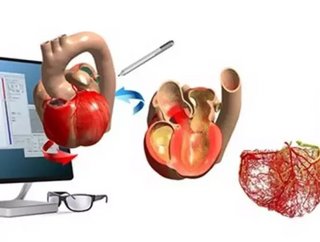
Fujitsu has announced that the University of Tokyo will use heart simulator viewers with stereoscopic displays compatible with virtual reality (VR) technology in an electrocardiogram lecture for third year students in the Faculty of Medicine. The simulator has been developed by both parties.
The heart viewer utilises as content the output data from a heart simulator jointly developed by Fujitsu and University of Tokyo using the K computer as well as a computer cluster.
Students will be able to see the complexity of "excitation propagation," a phenomenon whereby electric stimulation from pacemaker cells spreads throughout the heart, with a stereoscopic, 360-degree view utilising VR, allowing them to understand how electrocardiograms are created through this process.
Related stories
- New technology could revolutionise traditional vaccines
- New EndoBarrier tech looks set to rival traditional gastric bypass surgery
- How Philips has gone from electronics to transforming healthcare
Based on its use in the University of Tokyo's lecture, Fujitsu will proceed with the development of the heart viewer with an aim toward product commercialisation within Fujitsu's fiscal 2017 (ending March 31, 2018) for use as a more effective educational tool while contributing to the advancement of medicine.
By providing a stereoscopic view with VR, this technology supports the efficient teaching of medical students, enabling them to really see such factors as the interrelation between the graph shown on the electrocardiogram and the propagation of electrical signals, and the difference between the behaviours of the heart both in normal times and when diseased.
The technology accurately simulates the activity of the heart from the muscle cell level. As a result, it is now possible to use these 3D models to see not only the internal and external structure of the heart, but also such things as true-to-life heart muscle activity, detailed networks of blood vessels and the flow of blood, as well as the spread of electrical propagation.